MATERIAL AND TECHNICAL BASIS OF LABORATORY OF CLINICAL RECOGNITION
The laboratory of clinical diagnosis is equipped with the most modern medical equipment of the world famous American company "Beckman".
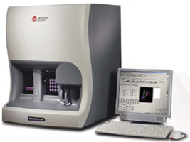 LH500 analyzer, intended for determining haematological analyzes, within 3 minutes perfectly identifies 27 sets of blood parameters with a laser method.
Microscopy of blood film Common urine analysis: Uriscan urine analyzer Common stool analysis
Microscopic investigation of smears of various kinds, 50 kinds of common infectious agents (chlamydia, mycoplasma, ureaplasma, trichomoniasis, syphilis, herpes, hepatitis, etc.).
Full microscopic analysis of urine, cerebrospinal fluid, sputum, prostatic fluid, duodenal contents and sperm.
Examination of the skin, nails and hair on pathogenic microbes.
Becman COULTER UNICEL DC600 biochemical device within an hour with the utmost precision determines 1,000 different analyzes.
• Biochemical tests – BECKMAN COULTER UNICEL 600
• Total protein
• Albumin
• Albuminuria
• Total bilirubin
• Bilirubin fractions (total, conjugated, unconjugated)
• Aspartate aminotransferase (AST)
• Alanine aminotransferase (ALT)
• Gamma glutaminetransferase (GGT)
• Lactic dehydrogenase (LDH)
• Thymol test
• Alkaline phosphatase
• Rapid glucose test (in capillary blood/ fingerstick glucose)
• Glucose in black blood
• HbA1C glycated hemoglobin
• Glucose tolerance test (sugar indication)
• Creatine clearance (Rehberg-Tareev sample)
• Urea, uric acid, creatinine and filtrate nitrogen in the blood
• Lipoprotein cholesterol (LDL), (HDL)
• Triglycerides
• Creatinine phosphokinase (CFK)
• Myocardial infraction of creatinine phosphokinase (CFK-MV)
• Amylase
• Diastase in the urine
• Na, K, Cl, Ca, Mg, Cu, P, Fe in blood
• C - reactive protein (CRP)
• Antistreptolysin O (ASLO)
• Rheumatoid factor (RF)
By means of the ACCESS 2 device of Beckman COULTER company we define infection and hormones with the modern methods
• FSH – follicle-stimulating hormoneCardiological tests
• Troponin I
• Myoglobine
• Myocardial infraction of creatinine phosphokinase (CFK-MV)
Tumor markers.
• Tumor markers of ovarium, glandula mammaria, gastrointestinal tract;
• PSA – prostate-specific antigen;
• SCC – squamous cell carcinoma;
• AFP – alpha-fetoprotein tumor marker
Identification of narcotic drugs in urine:
• Opiates
• Cocaine Metabolites
• Amphetamine and its derivatives
Infections
• Toxoplasma (IFA)
• Herpes – (IFA)
• Cytomegalovirus – (IFA)
• Chlamydiae – (IFA)
• Trichomonad – smear
• Mycoplasma – smear
• Ureoplasma – smear
• Gardnerella vaginalis – smear
• Syphilis – card test
• Rubella – (IFA)
• Brucella – (IFA)
• HBs Ag IFA
• HBs Ag test method;
• Hepatitis C test method;
• Hepatitis C IFA;
• Х- Pilori - card test;
• Х- Pilori IgM;
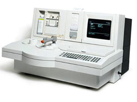 By means of the 200 ACL analyzer we define coagulation and blood clotting factor.
• Blood clotting time (by Sukharev)
• Prothrombin index and prothrombin time
• Fibrinogen
• Activated partial thromboplastin time (APTT)
• International normalized ratio (INR)
To carry out "Polymerase chain reactions" clinical diagnostic laboratory is provided with a fully automatic Rotor-Gene device from Switzerland
Polymerase chain reaction (PCR) - Rotor – Gene 6000 corbett
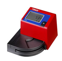 There are following devices in the clinical diagnostic laboratory of the hospital HemoCue WBC (white blood cell count rapid method), CEDY-40 (determination of erythrocyte sedimentation rate), Glukose (determination of the level of sugar in the blood) and HemoCue ALB (albumin determination in the urine).
As a result of measures taken in the direction of improving the technical capabilities of the clinical diagnostic laboratory, a complete blood count on 26 parameters, full laboratory study of blood of biochemical, hormonal and infectious natures, the study of common infectious agents in smears of different nature as a whole on 50 species (chlamydia , mycoplasma, ureaplasma, trichomoniasis, syphilis, herpes, hepatitis, etc.), a complete analysis of urine, cerebrospinal fluid, sputum, prostatic fluid, duodenal contents and sperm are conducted here.
As a result of the clinical diagnostic laboratory activity, here laboratory tests are carried out on a large scale, except for specific infectious diseases.
Şüa diaqnostikasi xidməti.
Maqnit Rezonans Tomoqrafiyası
This method is based on the measurement of the response to high-speed pulses of hydrogenic nuclei present in the tissue of stable magnetic field. MRI allows you to examine all the internal organs of the human body. 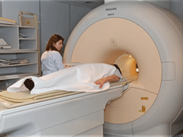 During the MRI patient is pulled into the cylinder, which is an electromagnetic field. The radio waves are sent to hydrogenic atoms of the body, making it possible for energy intake, the image comes to life on a computer screen and can be transferred to the X-ray film or photographic paper. MRI examination is completed between 20-40 minutes. MRI is used to evaluate the nervous system, bone marrow, muscle and joints and soft tissues. The advantage of this method lies in the fact that the patient is not exposed to radiation. The resulting image is considered to be clearer and more favorable from a diagnostic point of view.
MRI can detect changes in the brain, pituitary gland, eyeballs, neck, spine, spinal cord, organs of the chest, abdominal, pelvic organs, musculoskeletal system and soft tissues.
Computer Tomography
Computer tomography (CT) - a method of examination, based on the absorption by body tissues of varying degrees of X-rays. During a CT scan radiation source rotates around the table on which the patient lies, directed thin beam of rays passes through the object and is caught by a few X-ray detectors. In the process of imaging rays pass through the human body in several directions and are gathered by detectors. These detectors send this information to the computer, and the computer, in turn, depending on the degree of radiation absorption by tissues creates layering image and displays it on the monitor. Thus, registering the slightest changes in the absorption of rays it reflects them on the monitor that reveals small pathological processes.
CT scanner consists of a table and the round window (gantry) which table enters. On the surface of the window ring the radiation source (X-ray tube) is located on one side, and sensitive detectors on the opposite side. The patient lies on a table and the table is entered into a slowly revolving window. With one rotation of the window on the computer monitor slice of examined organ appears, measured in millimetres. Thus, a layered image of the body is received. The layer thickness is up to 0.3 mm, and receiving the slices takes 7-12 seconds.
In some cases: during the exam of vascular structures, tumors and their metastases, to obtain a more accurate image, and also during the evaluation of the functions of organs (liver, kidneys, etc) contrast agents may be used.
With this high-tech equipment timely and accurate diagnosis of central nervous system, chest organs, cardiovascular system, gastrointestinal tract, urogenital system, gynecological diseases, degenerative and destructive changes in blood vessels, spine, pelvis and the whole musculoskeletal system is conducted. CT is used to determine the localization and distribution of the pathological process in various organs and tissues of the body, to track the dynamics of various pathophysiological processes, select the approach and scope of surgery, perform a biopsy of tumors, evaluate the results of treatment.
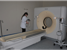 In early 2009, the most modern in the world CT scanner, manufactured by "Philips" was installed at the National Hospital (64-slice MDCT, multidetector or multislice computed tomography). The advantage of this device is that it allows the study of the heart. Previously, to visualize the state of the coronary arteries classic angiography was used - a method of inserting a catheter through the femoral artery (invasive). Despite informative, this method carries some risk and complications to the patient (hematoma, bruising, fibrillation). During the classical angiography cardio operation room must be in a ready state, so that in case of necessity it is possible to carry out the urgent surgical intervention. Then, for this study there is the need to transfer the patient to the hospital, to prepare him/her for observation and a few days after the observation to hold the the patient at the hospital. Now this process takes only 30 minutes and is performed on an outpatient basis.
The new scanner is equipped with a program "Package Heart View", provided for the study of the heart and coronary vessels. As this occurs: In order to determine the heart rate synchronized with the MRI on the body of the patient, special electrodes are fixed.
Thus, in order to avoid artifacts that occur when the heart contracts, the optimal scanner mode is selected. The production of contrast fluid through the automatic injector in the cubital vein is started and the program, identifying private hemodynamics of the patient, creates the protocol of scanning. After this the command is given to hold your breath for 10-12 seconds, and multislice tomography system starts to scan the heart, and in that time, the thickness of the cut is less than 1 mm. Thus, the procedure of the study is completed in 10-15 minutes. During scanning, a huge amount of information is obtained for processing. This powerful, most advanced software scanner provides a 3D (three-dimensional) image of the heart and coronary vessels, to determine the presence of stenosis and occlusions, find small growths of calcium in the blood vessels, to analyze the anatomical features of the heart.
The most important thing is that in this study there is no need to place the patient at the hospital and conduct preparatory work, without any risk or cause of deterioration.
This method is able to obtain a comprehensive screening of coronary heart disease. It is indispensable for children's pathologies associated with a greater risk of catheterization. A major factor is that now everyone can explore the heart without a long arduous process of preparing and fear. This fear prevents the patient from seeing doctor in time, which subsequently leads to a deterioration in the next stages.
In addition to the above, this scanner was equipped with a number of unique programs which give volume, 3D (three-dimensional) image of all human anatomical structures, a special program "Dental", indispensable in dentistry and implantology, a measurement of the liver which is an extremely informative study of the density of the liver tissue and liver transplantation, and 3 phase (arterial, venous and parenchymal) dynamic research of the organs.
Interested parties may contact on all matters the radiology department of the Republican Hospital named A.N.Heydarov of MIA.
X-Ray
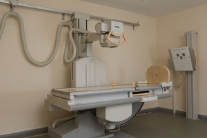 X-ray examinations are carried out by a digital device "DuoDiognost Philips".
Physiotherapeutic Treatment
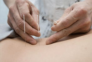 TTreatment of various diseases using natural factors begins with the creation history of mankind. Historical sources show that, even before B.C., ancient people treated some diseases, bodily injuries with hot and cold compresses, but the skin and gastrointestinal diseases with washing and clysters. Using the elements of treatment methods such as manual therapy and acupuncture and applying the inhalation of musculoskeletal system, nervous system and a variety of medicinal herbs, our great ancestors treated respiratory tract diseases.
Hippocrates who said: “Nature is the doctor of diseases”, widely used sun baths, hot and cold water procedures, massage, therapeutic exercise and climate. The ancient Chinese doctors accelerated the healing of diseases making different slashes on the skin, puncturing, applying the lit cigars to the different parts of the body.
Over the past decade, physical methods with pathogenetic effect have been used widely in the developed countries of the world for treatment, rehabilitation and prophylactic purposes.
The interest has been increased in the application of natural treatment factors due to certain difficulties and side effects of use of the pharmacotherapy which is a traditional method of treatment. Treatment with physical factors is more favorable in terms of finance, so it costs much cheaper. On the other hand, in some cases, complex use of the drugs allows to reduce the duration of treatment. The Physiotherapy Department of Republic Hospital of the Ministry of Internal Affairs named after A.Heydarov has been supplied with the newest physiotherapy equipments of various countries (Germany, Italy, Russia, and Turkey) of the world.
Each of these equipments consists of apparatuses regulated and combined by modern computer system related to the scientific - technical progress.
It is possible to perform the following physiotherapeutic treatments by the help of these apparatuses:
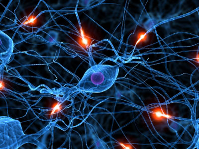 Bed fund of the Department of Neurology has 40 beds.
Almost 12-15% of admitted patients during a year fall to the share of the Department of Neurology.
Fast and hard lifestyle of the modern period shows that neurological diseases are also one of the actual problems for the protection of health willing or not. Besides Chief of the Department, 5 physicians work in the Department both civil and ranked.
Educated and professional medical staff that have taken practical course in the advanced clinics of Azerbaijan, Russia, Turkey and Europe stands resolutely on guard of health of the employees of the Ministry of Internal Affairs, their family members and pensioners or other contingent.
Work of careful, responsible nurses is obvious case of an irreplaceable role of them.
Endoscopic urology
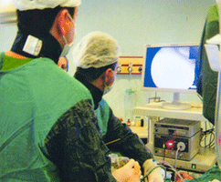 Endoscopic examination and surgical services in Urology in comparison with other areas of medical science began quickly and achieved great success. Bond of most urological organs among themselves in different ways in urology greatly facilitates the implementation of endoscopy. Today in modern clinics of developed countries urology is composed of endoscopic surgical service. It became possible by endoscopic way to reach any organ of urinary system, conduct the operation.
Uronephrological Department of Republican Hospital named after A.N.Heydarov of the Ministry of Internal Affairs was formed as a department, consisting of the endoscopy cabinet, equipped with the latest equipment, dressing room, department of distance shockwave lithotripsy, urological operating room meeting the European standards, and the availability of about 20 hospital beds. Doctors of the department are modern professionals with scientific degree, qualified in urological schools of Russia-Turkey-Europe with 15-20-30 years of experience. Three doctors of the department are members of the European Society of Urology.
Urological services currently provided by physicians of the department:
1. Open surgery
Surgical operations in the kidney and urachus (stone, tumor, plastic surgery)
Tumors, stones, bladder diverticula
Prostatic hyperplasia
Operations on the scrotum (hydrocele, varicocele, orchiectomy, orchidopexy)
Hypospadias, circumcision
Inguinoscrotal hernia
Incisional hernia
2. Endoscopic intervention
Cystoscopy, urethrocystoscopy
Closed surgery of the prostate (TUR, bipolar, monopolar)
Cystolithotripsy
Endoscopic lithotripsy of urachus
Ureterorenoscopy
Installation and removal of the Double J catheter
Treatment of the narrowness of the urethra
Retrograde intrarenal surgery
Percutaneous nephrolitholapaxy
3. Male infertility
USD of male genital organs
Laboratory testing of urine, semen, prostatic fluid
Determination of sex hormones
Pathogenetic medical and physiotherapy treatment of the processes of chronic inflammation of the prostate, seminal sacs and scrotum (magnetic-infrared-laser treatment, interferent therapy, pneumomassage, electrophoresis, phonophoresis, rectal microwave therapy, cryotherapy, etc.)
Diagnosis and pathogenetic treatment of impotence
Transrectal puncture of the cyst, abscess of the prostate
4. Diagnosis of the uronephrological patients (clinical-laboratory-instrumental) and pathogenetic treatment (medicamentous and physical therapy in a wide aspect)
In recent years, doctors of Urology Department of the Hospital conducted about 1,000 endoscopic urological surgical operations.
 The bed space of the Department of Cardiology consists of 56 beds.
Approximately 17-20 % of patients admitted to hospital during the year account for the Department of Cardiology.
The development of the cardiology with the most morality rate in the word statistics proves once again that which weight that field owns in the general health protection.
Besides the Department Head, 5 physicians including civil and ranked work at the division.
The educated and skilled medical personnel, who have completed the course of practice in the leading clinics of Azerbaijan, Russia, Turkey and Europe, worked determinedly for the health of the employees of Ministry of Internal Affairs, their family members and pensioners as well as the other contingent. The works of caring and responsible nurses are an obvious example which once more proves their significant role in this department.
 Endokrinologiya şöbəsinin çarpayı fondu 24 çarpayılıqdır.
İl ərzində hospitala qəbul edilən xəstələrin təqribən 7-8 %-i endokrinologiya şöbəsinin payına düşür.
Dünyanın “pandemiyası” kimi qəbul edilmiş metabolitik sindrom və şəkərli diabet xəstəliyi bu sahənin yüksək səviyyədə inkişaf etdirilməsi zərurətini yaradır. Son zamanlar qalxanabənzər vəz və digər endokrinoloji xəstəliklərin sürətlə artması endokrinologiya şöbəsinin də işinin nə qədər məsuliyyətli olduğunu bir daha sübut edir.
Şöbədə həm mülki, həm də rütbəli olmaqla şöbə müdirindən əlavə 3 həkim fəaliyyət göstərir. Azərbaycanın, Rusiya, Türkiyə və Avropanın qabaqcıl klinikalarında təcrübə kursu keçmiş savadlı, bacarıqlı həkim kollektivi istər Daxili İşlər Nazirliyinin əməkdaşları, onların ailə üzvləri və təqaüdçülərinin, istərsə də digər kontingentin sağlamlığının keşiyində əzmlə dayanmışlar. Qayğıkeş, məsuliyyətli tibb bacılarının əməyi də bu şöbədə onların bir daha əvəzsiz rol oynadıqlarının bariz nümunəsidir.
 Bedspace of the therapeutic department consists of 65 beds.
Approximately 20-25% of patients, admitted to the hospital within a year, fall to the department of therapy.
The department is provided with medicines and equipment that meet modern standards.
In addition to the chief, there are 6 doctors in the department, both civilian and military. Educated, skilled team of doctors, completing training courses in leading clinics of Azerbaijan, Russia, Turkey and Europe, stands firmly on guard of health both members of the MIA, their family members and pensioners, and another contingent.
The work of caring, responsible nurses is also a good example of their irreplaceable role in this department.
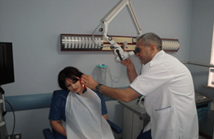 The Department of Otorhinolaryngology of Republican Hospital of MIA named after A. Heydarov is provided with medical instruments, equipments and devices meeting modern standards, and only manufactured in Europe.
As a result, examination and treatment of ear, nose and throat diseases is carried out more effectively, quickly, convenient and comfortable for the patient.
ENT unit made by EUROCLINIC in Italy, in the Endoscopic examination room, allows observing pathological changes in the ear, nose, throat and vocal cords on the monitor.
Therefore, the image is transmitted via camera to the monitor with 0, 30 and 70 degree rigid endoscopes. Above mentioned different degree rigid endoscopes create an opportunity to observe fine structures of ENT parts under 0, 30 and 70 degree angles.
Moreover, beside viewing of nose, nasopharynx, pharynx and larynx on the monitor, flexible endoscope of German ENT endoscope Karl Storz, gives an opportunity to remove foreign bodies (fish bone, etc.) from these organs with special gripper through the working channel.
About the Department of Surgery of the Hospita.
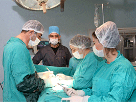 The Department of Surgery of the Hospital takes a leading position due to performing surgical operations of various complexities in Azerbaijan. Surgeons working in the department participated in the qualification improvement courses and they perfectly carry out endoscopic as well as voluminous open surgical operations in the abdominal cavity and thorax. The Department of Surgery consists of one Honoured Doctor of Azerbaijan Republic, Candidate of Medical Sciences and physicians with high category and scientific potential. Each year, up to 1000 surgeries are performed in different directions on 10 profiles in the Department of Surgery of the hospital: abdominal, thoracic, gynaecological surgeries are carried out, neurosurgery, modern reconstructive technologies are applied. Modern therapeutic and diagnostic equipments such as Ethicon, Olympus, in the department of surgery and surgical unit which were produced by the medical equipment manufacturers allow carrying out surgical operations successfully.
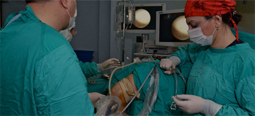
Open and laparoscopic surgical operations conducted in the department of surgery.
The Department of Ophthalmology of the Hospital is provided with up-to-date equipment.
Latest model equipment of “TOPCON” Company of Japan has a great role in diagnostics, prevention and treatment of eye diseases.
Multifunctional “FOROPTER” enables eyesight test and glasses assignment.
Biomicroscope enables to determine anterior and posterior segment of an eye.
Avtoreftometr measures eye refraction and assigns exact glasses.
Pneumotonometer enables noncontact measurement of intraocular pressure.
Fundus of eye is examined using 78 dpt aspheric lens of “VOLK” Company and ophthalmoscope of “Reister” Company of Germany.
The latest method of cataract surgery is available using “LAUREAT” phacoemulsifier of “ALKON” Company.
Eye exam – is subsequent exams in order to determine vision.
Level of vision should be determined initially in order to conduct exam correctly.
One of the first complications of eye diseases is eyesight weakness.
Exam of normal eye consists of 4 (four) steps:
1. Examination of visual acuity and refraction;
2. Examination of anterior segment using biomicroscope
3. Examination of fundus of eye
4. Ocular tension measurement
1. Myopia, hypermetropy and astigmatism are the main causes of eyesight weakness. Sometimes these eye disorders are available in both eyes, as well as one of eyes can be healthy, but another one can be defective. But there may be different defects in both eyes. Eye defects vary according to the age. Main purpose of eye exam is not only for determination of eye defects, but also use of correct treatment method. (glasses, contact lens, surgery or laser surgery)
2. Assessment of the structures such as anterior lids, eyelashes, conjuctiva, cornea, sclera, iris, eyeball, crystalline lens, vitreum in the biomicroscopic examination. Eye complications should be paid attention. Prevention of severe eye problems is possible with early intervention.
3. Eye fundus (retina vessel and nerve) exam is carried out after waiting for 30 minutes approximately from squeezing drops to dilate pupils.
4. Regular ocular tension measurement is necessary.
Even if the patient has no complications, only with eye exam it is possible to define hidden problems.
Glaucoma
Glaucoma (is known as ocular tension among people). This disease mainly begins after 40 years and increases during years and damages optic nerve that is important for vision. Loss of vision cannot be recovered during glaucoma. Therefore, early detection and treatment of this disease is very important. Careful eye examination is a main condition for early detection of this disease.
What is cataract?
Cataract is loss of transparency of natural crystalline lens accumulating beam. Crystalline lens losing transparency becomes as frosted glass and eyesight gets weakened. Eye fatigue and headache are on of the characteristic complaints alongside with nonfocal and blurred vision. Cataracts are very common in older people. However, it can be formed in newborns, diabetic patient after intraocular inflammation. Stop of cataract increase or recovery is impossible with treatment (without surgery).
The only method of cataract treatment is surgical. Cataracted crystalline lens is removed from eye and transparent lens is placed instead of it. Stitching had been used for elimination of cataract before. Such a surgery lasted more and there were complaints as sticking, burn and blurred vision after surgery. Stitches are needed to be unripped in this method. But nowadays modern methods are improved.
As the most modern cataract surgery, in phacoemulsification intraocular section is entered from a very short excision cornea with an instrument and crystalline lens is divided into a very small pieces by ultrasonic energy and taken by vacuum. Lens according to eye is placed from this excision and surgery is completed without stitching. Phacoemulsification is a very reliable and less complicated method increasing vision level in the earlier period as it is carried out by a little excision and closed system. The stitch is not unripped after surgery.
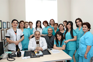
The solution for the chronic renal failure and hemodialysis problems was started at Republican Clinical Urology Hospital by forming "Kidney transplant and hemodialysis department" (Kidney Center) in 1970 (Sultan Aliyev was assigned as the first head of the department). The first hemodialysis was successfully carried out in September, 1970. A wide range of clinical, scientific and practical activities was implemented thanks to great efforts under the guidance of academic M. Javad-zade.
Orqanizmin həyat fəaliyyəti nəticəsində insanın orqan və sistemlərinin işinə və funksional vəziyyətinə mənfi təsir göstərə bilən xeyli miqdarda toksik maddələr yaranır və ya The solution for the chronic renal failure and hemodialysis problems was started at Republican Clinical Urology Hospital by forming "Kidney transplant and hemodialysis department" (Kidney Center) in 1970 (Sultan Aliyev was assigned as the first head of the department). The first hemodialysis was successfully carried out in September, 1970. A wide range of clinical, scientific and practical activities was implemented thanks to great efforts under the guidance of academic M. Javad-zade.
 Of course, human organism has disposal systems which neutralize toxins and self-defense mechanisms against the harmful factors. However, the bodies and mechanisms with the function of cleaning the organism from "toxins" cannot always cope with this work due to the urbanization, spoiled ecology and fast life rhythms. This leads to the accumulation of unnecessary agents and changes in the internal environment of the body, so serious metabolic disorders and pathological cases, which are dangerous for the organism, occur. Thus, a number of diseases based on the pathological changes in the immune system (allergic changes, autoimmune reactions, the weakening of the immune system and resistance) and disruption of metabolic processes, such as hypertension, ischemic heart disease, sclerotic changes in vessels of the brain, diabetes, gout, etc. develop.
The traditional treatment methods applied in most of these diseases only eliminate the symptoms of the disease, - prevent the development of allergic reaction or infection, dilate the bronchi during bronchial asthma or lower blood pressure, etc. At the same time, it does not affect to the main cause of the disease and is achieved at the expense of a number of side effects.
As a logical conclusion it may be assumed that the excretion of the toxic substances which are the products of organism’s activity can be a turning point in the course of disease. In this view, using the extracorporeal hemocorrection methods it is possible to obtain satisfactory results during the treatment process.
Extracorporeal hemocorrection is the quality and quantity change of the blood cell elements, re-development of the protein and water-electrolyte, enzyme and gas composition with a variety of technical methods outside the organism. Hemocorrection is conducted through the efferent (efference-excretion) therapy methods. Efferent therapy methods- direct removal of harmful substances got to the organism at the background of various diseases or from the external environment have been directed to the internal purification of the internal milieu.
The most effective types of these methods which are used more in clinical practice are the following:
- Hemodialysis;
- Hemosorption;
- Plasmapheresis;
In this case, toxins, nitrogen byproducts (creatinine, urea etc.), immune complexes, cholesterol and other pathological products causing a variety of diseases are removed from the blood. Selective effects such as detoxication (purification of the toxic substances from the organism), reocorrection (improve the rheological properties of the blood), immunocorrective (boost the immunity) stand on the basis of each method.
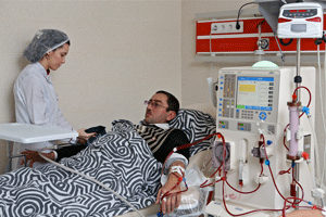
I. Hemodialysis (hemo-blood, dialysis-purification) is a method in which the blood is removed from the body and purified passing through the hemofilter (dialyser) and the purified blood returns to the organism. Hemodialysis is carried out through the “artificial kidney” device.
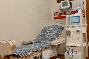
Hemodialysis room and osmosis-water treatment room of Republican MIA Hospital named after A.Heydarov.
The device works on the principle of blood treatment by transferring the semiconductor membrane-dialyzator (hemofiltration) made of special material and enabling to perform certain functions of the human kidney (removal of nitrogen residues, water-salt exchange, regulation of homeostasis and etc.) and the osmotically active dialysate of special compound and adapted to the gas and electrolyte composition of the blood (the buffer solution in accordance with the blood composition-acetate and bicarbonate).The device is combined with the blood circulation by puncturing the special vascular access created with the help of surgical operation using the fistula needle (fistula). The blood enters the dialyzer through the magistrial shunts for purification. At this time, a certain part of the blood (200-300 ml) remains outside the body, circulates uninterruptedly, is purified from hazardous substances and eventually returns to the blood circulation of the body. As a standard the dialysis program usually takes 3 -4 hours a week and this program is called hemodialysis or periodic hemodialysis and chronic hemodialysis. The majority of patients come to special departments - hemodialysis centers 3 times a week and receives this operation on an outpatient basis. If a patient is selected for hemodialysis under the proper instruction, he (she) can receive treatment for years or 10 years and lives an idyllic life by the help of dialysis.
There are two types of hemodialysis:
- Acute hemodialysis
- Chronic (program) hemodialysis
Acute hemodialysis is applied during the acute renal failure caused by a variety of etiologic factors. After a few hemodialysis session a patient returns to normal life and if serious irreversible pathology occurs at the level of the kidneys, the permanent chronic hemodialysis will be continued in accordance with the living index or a patient will be sent to the renal transplantation.
Tips for Chronic (software) hemodialysis (during Chronic Renal Failure):
1. Renal clearance of creatinine 10-15 ml/min/less than 1,73 m2;
2. Renal clearance of urea 7 ml/min/less than1,73 m2;
3. Creatinine level in the blood - higher than 600-700 mk mol/l;
4. Potassium level in the blood-higher than 6,5 mmol/l;
The presence of one of the following clinical signs:
- symptoms of uremia;
- hyperhydradation, uncontrolled AH, congestive heart failure, starting pulmonary edema;
-progressing uremic neuropathy;
- metabolic acidosis, in the case of decompensation;
-uremic pericarditis, uremic precoma;
- protein–energy malnutrition, hypercatabolism.
Relative contra-indications against the program (chronic) hemodialysis:
1. Malignant tumor;
2. Ischemic heart disease, myocardial infarction, heart failure;
3. Decompensation phase of chronic hepatitis, cirrhosis of the liver;
4. Obstructive lung diseases, active TB;
5. Damage of cerebral vessels;
6. Mental diseases (schizophrenia, epilepsy, psychosis etc.)
7. Blood diseases (aplastic anemia, leukemis etc.)
8. Low intelligence level, the lack of interest in social rehabilitation (alcoholic, drug addict, homeless hobo);
9. Severe diseases of the nervous system;
10. Patients with a high risk of massive bleeding in the case of anticoagulant injections. (uterus fibroid, Mallory–Weiss syndrome, stomach and Duodenal ulcer). Aneurysm of the heart and magistral arteries.
 It should be noted that it is impossible to guarantee for the help of hemodialysis treatment for patients when the clinical course of the disease gets worse as a result of not referring the patients with chronic renal insufficiency and potentially in need of the program hemodialysis to a nephrologist on time.
For that reason, timely sending of a patient to nephrologist for advice is very necessary as measures taken with these specialists allows to remove the CRF complications in pre-dialysis period. On the other hand the improper specification of instructions for hemodialysis treatment leads to further deterioration of the patient's condition and acceleration of fatal ending.
In general, hemodialysis is not only the treatment but also a way of life!
Baku city, 36 Ziya Bunyadov avenue. Zip/postal code: AZ1069
E-mail: hospital@mia.gov.az